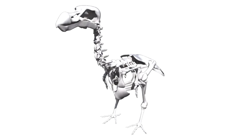
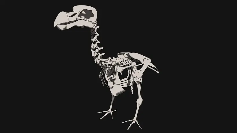
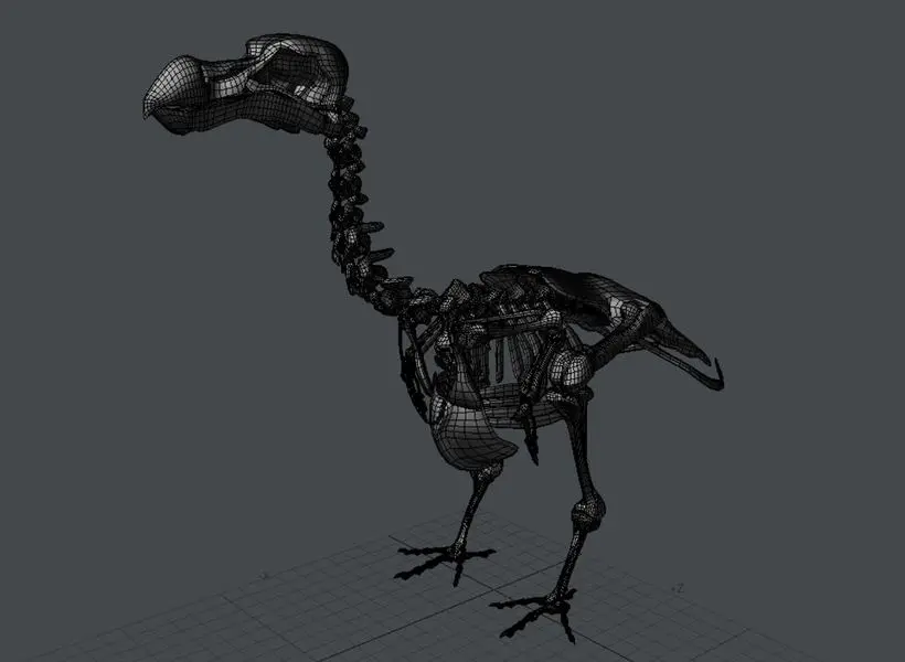
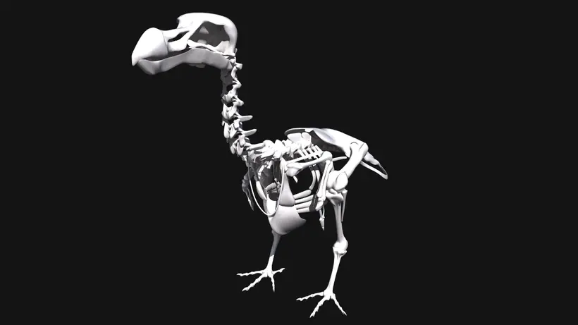
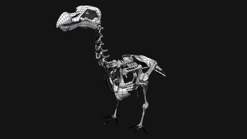
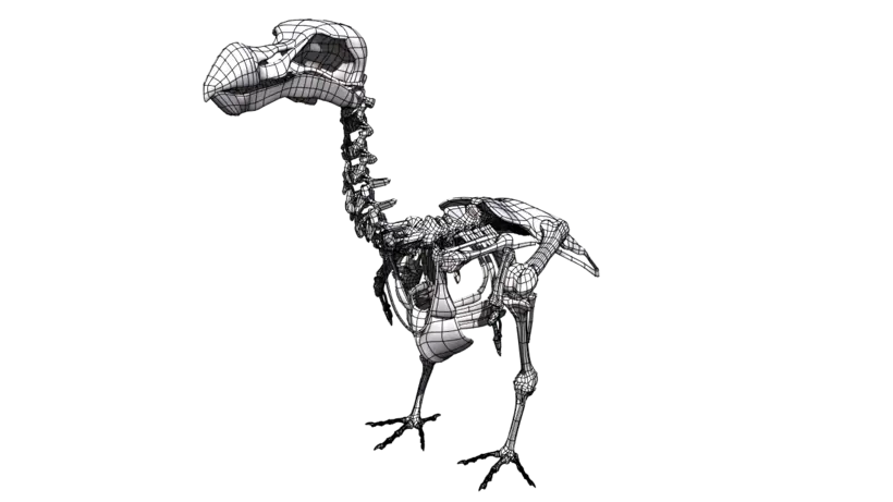
Modelled on a decimated 3-D scan of the Thirioux dodo skeleton on display at the Durban Natural Science Museum in South Africa. The skeleton has been digitally remounted in an anatomically correct pose. The stance has been lowered and the angle between the pelvis and the ribcage skeleton made less acute, amongst others. The original specimen is the subject of a newly published anatomical atlas of the dodo (Raphus cucullatus), the first major revision of dodo anatomy since 1866.
Free File Conversions: Need this 3d model in another format? please Contact Support to verify we can provide the format you need.
Please use the Support Forum if you have any questions or problems.
Please rate and review in the Review section.
Please rate and review in the Review section.